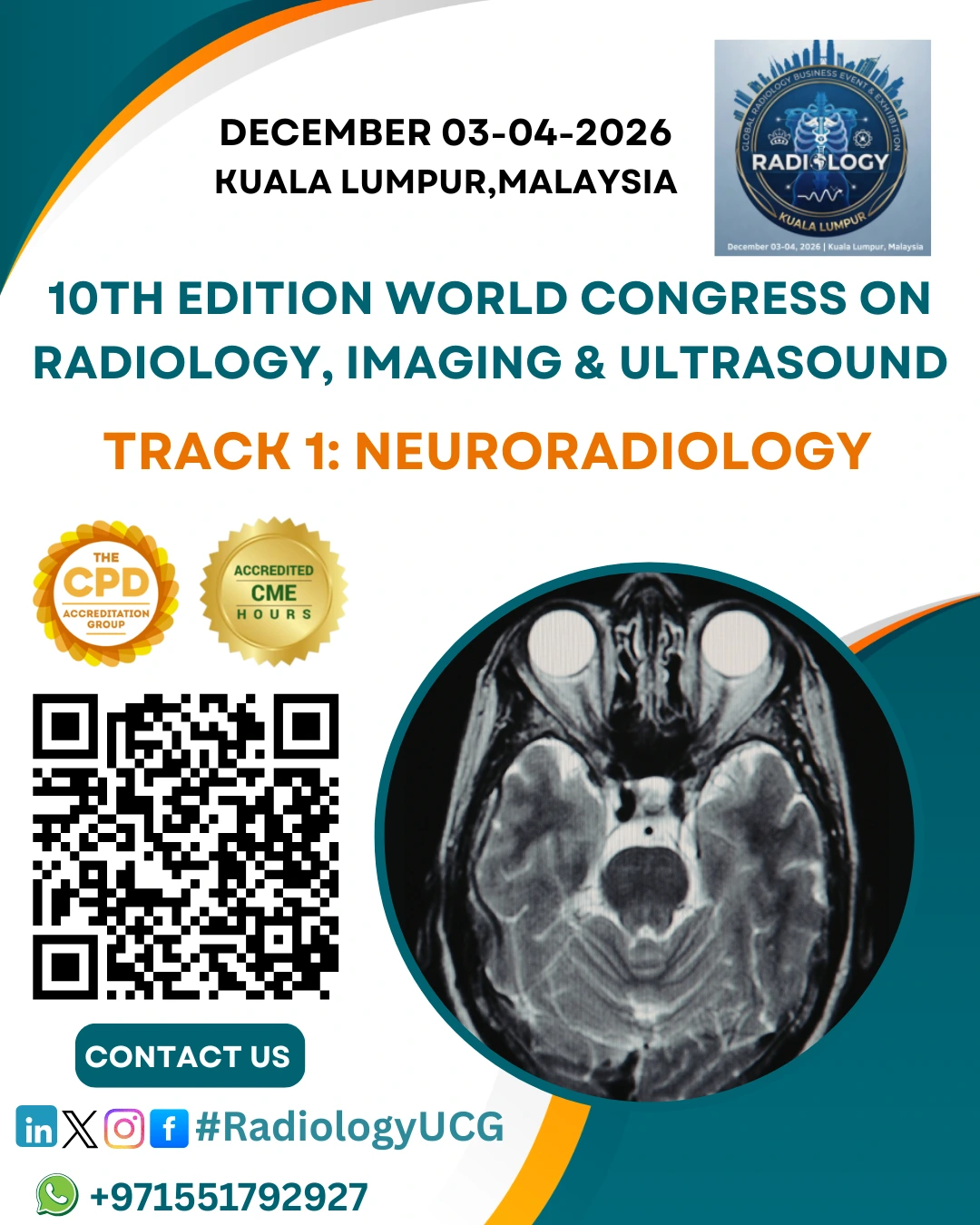
Sub Topic: Neuroradiology
Neuroradiology is a specialized branch of Diagnostic Radiology that...
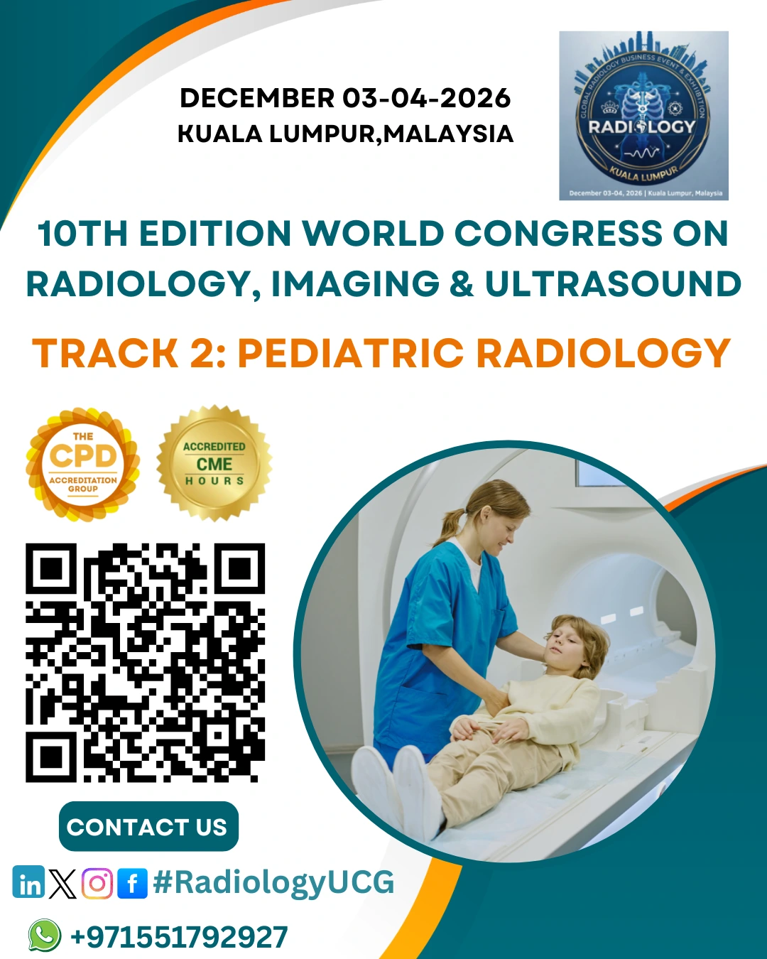
Sub Tracks: Pediatric Radiology
Pediatric Radiology is a specialized branch of radiology...
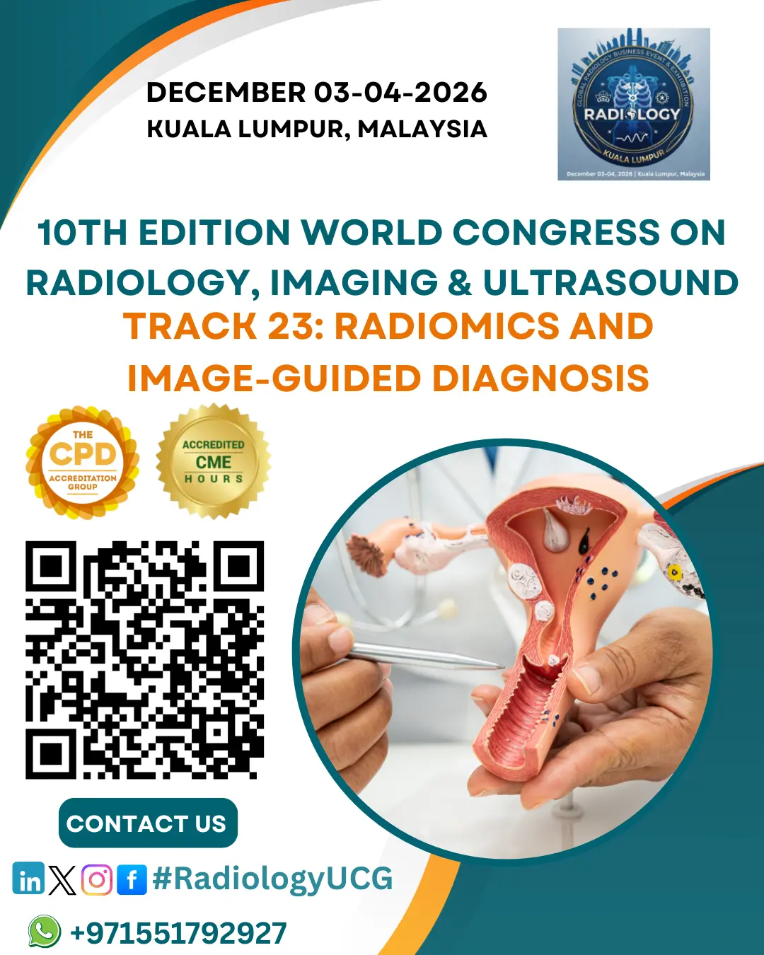
Radiomics is the process of extracting large amounts of quantitative data from medical imaging modalities such as CT, MRI, PET, and Ultrasound. These data include measurements of texture, shape, intensity, and tissue patterns—many of which are not visible to the human eye. By converting images into measurable data, radiomics provides a deeper understanding of disease characteristics.
Image-guided diagnosis uses real-time imaging to detect and evaluate diseases. When combined with clinical information, laboratory results, and genetic markers, radiomics enables more accurate diagnosis, prognosis prediction, and personalized treatment planning.