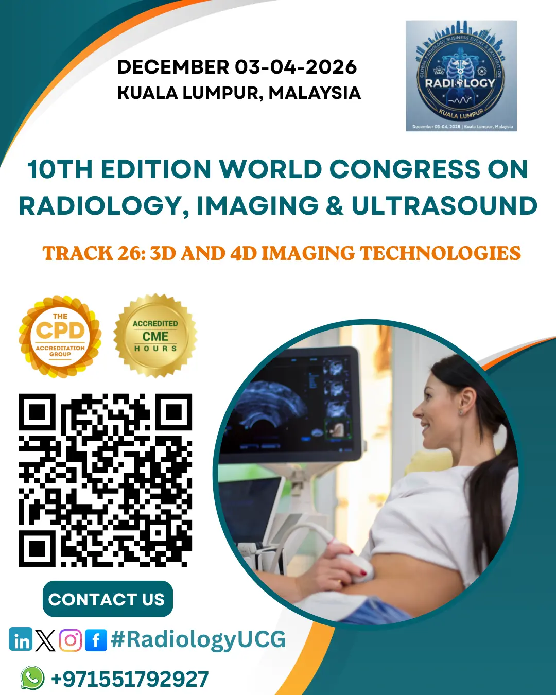
3D and 4D Imaging Technologies
3D and 4D imaging technologies are advanced medical tools that provide detailed, realistic views of internal body structures, enhancing diagnosis and treatment planning.
3D Imaging
-
Combines multiple 2D images from different angles to create a three-dimensional representation.
-
Allows precise examination of shape, depth, and spatial relationships of organs and tissues.
-
Common techniques: 3D ultrasound, 3D CT, 3D MRI, and 3D mammography.
-
Applications: obstetrics, cardiology, oncology, neurology, and orthopedics, aiding in diagnosis, surgical planning, and patient monitoring.
4D Imaging
-
Adds the dimension of time to 3D imaging, producing real-time moving images.
-
Enables observation of physiological processes as they occur.
-
Examples: 4D obstetric ultrasound for fetal movement, 4D cardiac imaging for heart function, and 4D imaging in radiation therapy for tracking tumor motion during breathing.
Benefits
-
Enhances diagnostic confidence and patient education.
-
Supports minimally invasive procedures.
-
Provides clearer, dynamic visualization for safer, more precise, patient-focused care.
While specialized equipment and trained personnel are required, the advantages of 3D and 4D imaging technologies make them essential in modern healthcare.