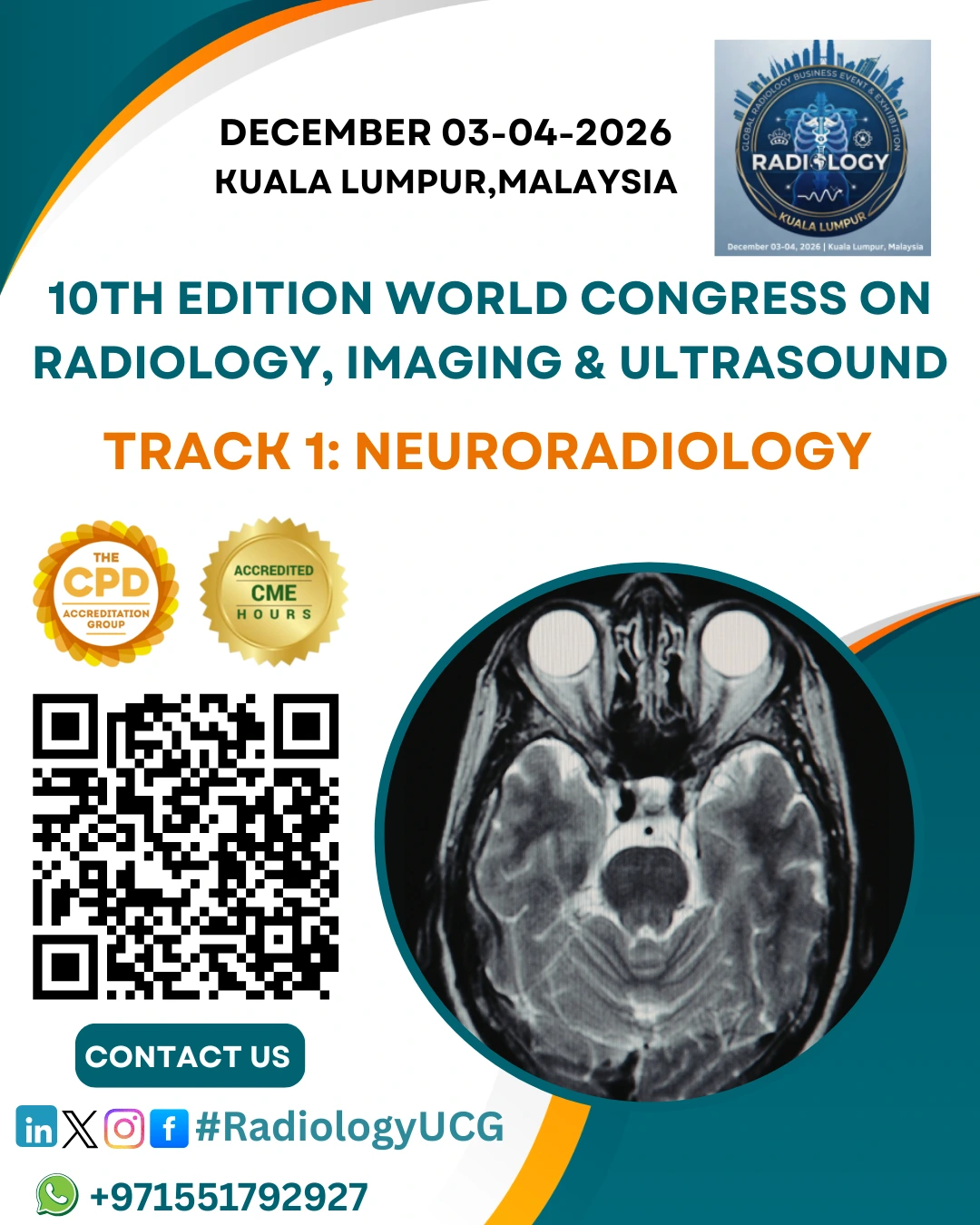
Sub Topic: Neuroradiology
Neuroradiology is a specialized branch of Diagnostic Radiology that...
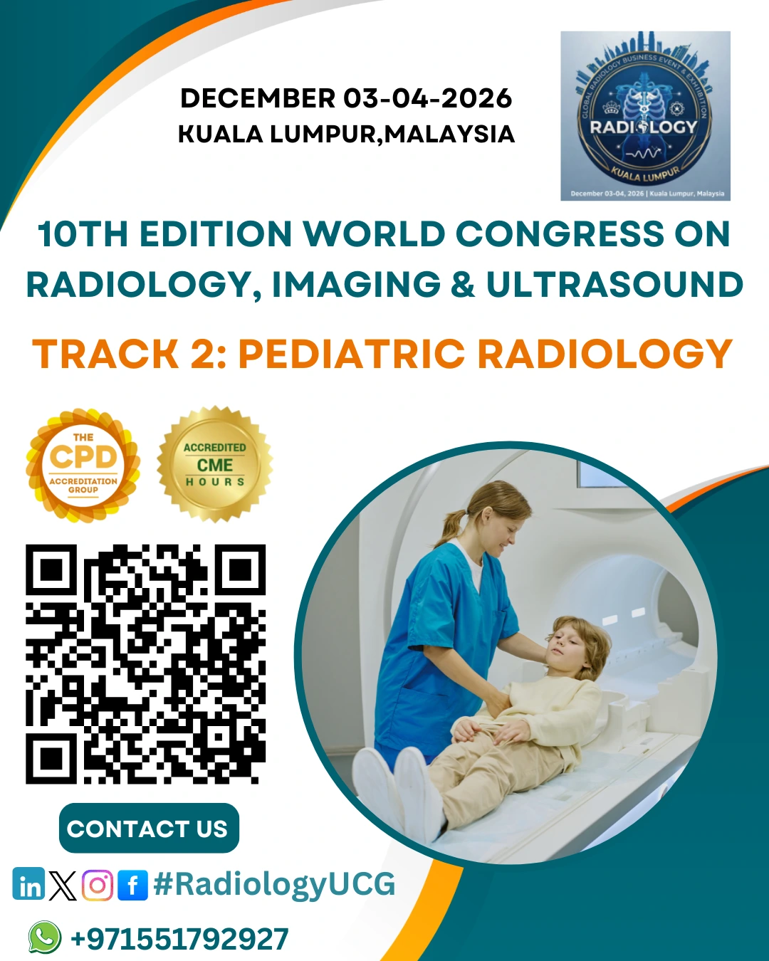
Sub Tracks: Pediatric Radiology
Pediatric Radiology is a specialized branch of radiology...
Sub-Topics in Chest and Thoracic Imaging
Advanced CT Techniques (e.g., HRCT, Dual-Energy CT, CT Pulmonary Angiography)
Thoracic Ultrasound & Image-Guided Interventions
Pulmonary Oncology Imaging
Interstitial Lung Disease (ILD) Imaging
Pediatric Thoracic Disorders
Pleural and Mediastinal Imaging
Airway and Obstructive Lung Disease Assessment
Emergency and Critical Care Thoracic Imaging
Cardiothoracic Imaging (Heart and Great Vessels)
Image-Guided Thoracic Procedures (Biopsy, Drainage, Ablation)
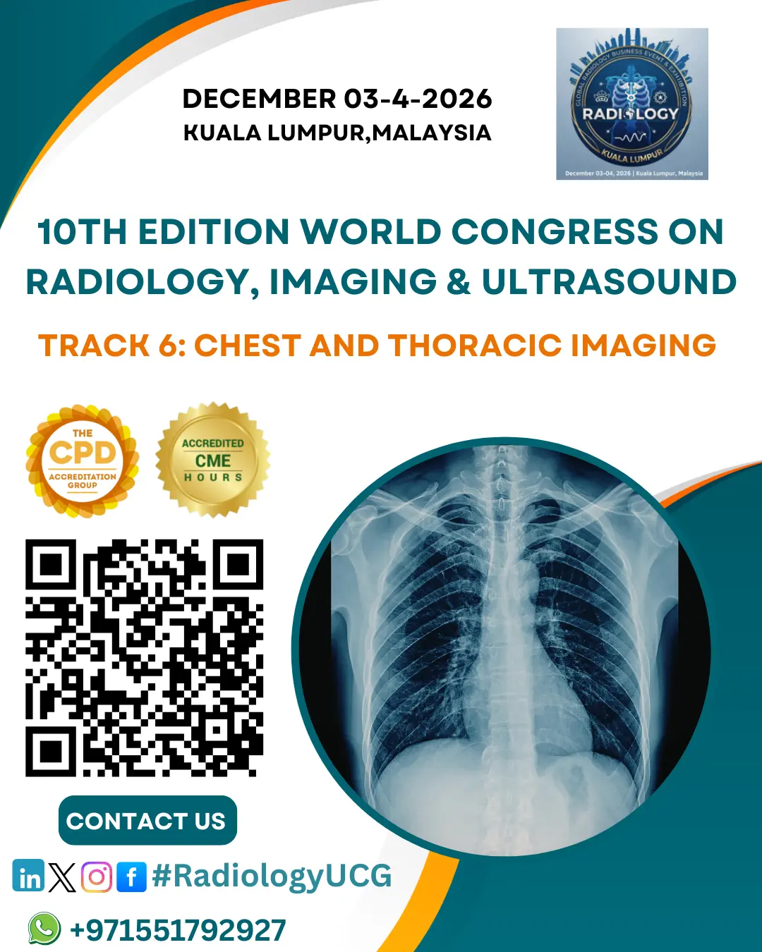
Overview of Chest and Thoracic Imaging
Chest and Thoracic Imaging is a specialized area of diagnostic radiology focused on diseases of the lungs, airways, pleura, mediastinum, heart, and thoracic vessels. With the global burden of respiratory diseases, lung cancer, infections, and cardiovascular conditions, the importance of accurate thoracic imaging continues to increase.
This specialty utilizes multiple imaging modalities—including Chest X-ray, CT, High-Resolution CT (HRCT), MRI, Ultrasound, and PET-CT—to provide precise diagnosis and guide treatment decisions. Recent advancements such as AI-assisted detection, low-dose CT for lung cancer screening, and functional imaging techniques have significantly enhanced diagnostic accuracy and early disease detection.
Core Focus Areas