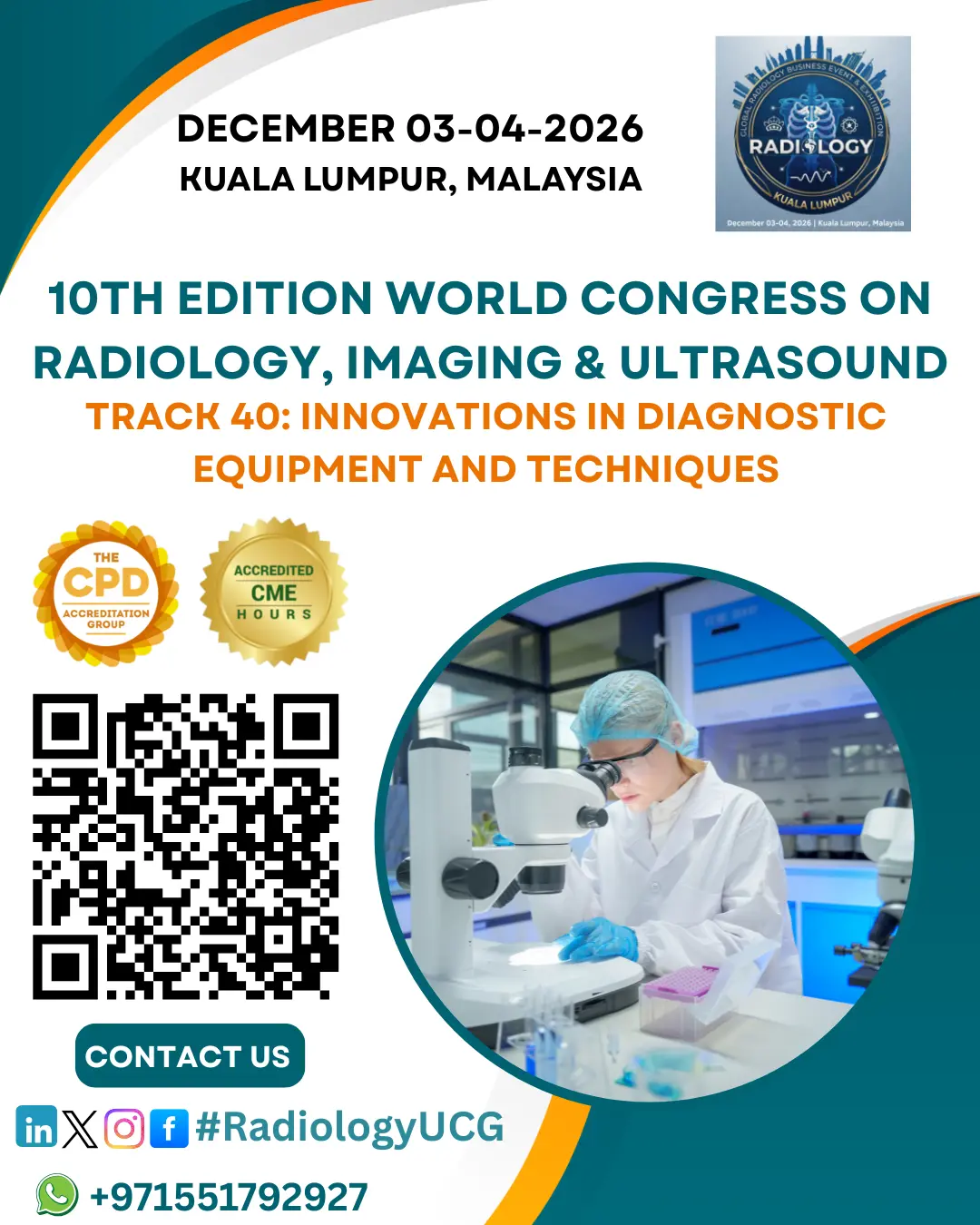
Innovations in Diagnostic Equipment and Techniques
Accurate diagnosis and precise planning are critical in neurosurgery due to the complexity of the nervous system. Recent advancements in diagnostic equipment and imaging techniques have significantly enhanced the ability to visualize, assess, and treat neurological conditions.
Advanced Imaging Technologies
-
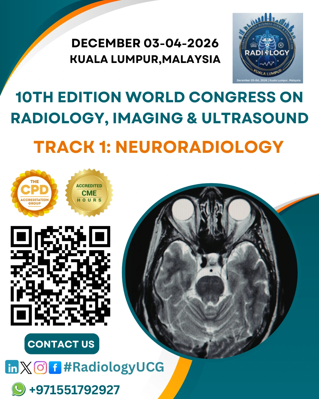
High-Resolution MRI: Provides detailed images of the brain and spinal cord. Functional MRI (fMRI) maps areas related to motor, sensory, and language functions, while Diffusion Tensor Imaging (DTI) tracks neural pathways for safer tumor or epilepsy surgeries.
-
Enhanced CT Scans: Rapid, high-resolution CT imaging allows quick evaluation in trauma cases. 3D CT angiography visualizes blood vessels, aiding neurovascular procedures such as aneurysm repair.
-
Positron Emission Tomography (PET): Offers metabolic and molecular imaging to distinguish between tumor recurrence and post-surgical changes, guiding targeted therapies.
-
Intraoperative Imaging: Tools like intraoperative MRI (iMRI) and CT provide real-time imaging during surgery to confirm tumor removal or implant placement. Ultrasound enables real-time guidance for tumor localization or cyst drainage.
Minimally Invasive and Image-Guided Techniques
-
Neuronavigation Systems: Function as a “GPS” for the brain and spine, using preoperative imaging to guide surgical instruments with precision and minimize risk to vital structures.
-
Stereotactic Biopsy: Combines CT or MRI with a stereotactic frame for precise, minimally invasive targeting of deep brain lesions.
-
Endoscopic Imaging: Miniaturized cameras allow visualization of intracranial structures through small openings, reducing tissue trauma and speeding recovery.
-
Optical and Molecular Imaging: Fluorescence-guided surgery highlights tumor margins, while molecular imaging detects abnormal tissue at a cellular level.
Neurophysiological Monitoring
-
Intraoperative Electrophysiology: Utilizes EEG, EMG, and evoked potentials to monitor brain and spinal cord function during surgery, allowing immediate correction of nerve or spinal injury.
-
Brain Mapping: Identifies essential functional areas during awake craniotomy, preserving speech, motor, and sensory abilities while removing lesions.
Artificial Intelligence and Machine Learning
-
AI-Assisted Imaging: Automates image analysis to detect subtle abnormalities, aiding in tumor segmentation and surgical planning.
-
Predictive Analytics: Integrates imaging, genetic, and clinical data to predict surgical risks and optimize personalized treatment strategies.